
Wrist radiography for hand bone age tells a lot; a girl with SHOX deficiency
 Abstract views: 478
DOI:
https://doi.org/10.51271/jpea-2022-194Keywords:
SHOX Deficiency, Madelung Deformity, Radiography, Short statureAbstract
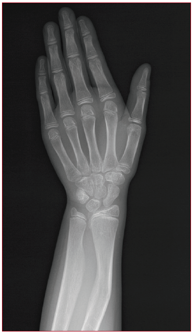
Madelung's deformity (MD) occurs as a result of premature closure of the medial and volar aspects of the distal radial physis.1 It is more frequent and severe in girls, and usually develops in middle/late childhood.2 MD is one of the most characteristic features of he short-stature homeobox gene (SHOX) deficiency, which causes short stature3. Radial bowing is one of the well-known radiological futures. On the other hand, there are three typical radiological sign of the hand radiograph for SHOX deficiency; triangularization, pyramidalization of the os lunatum, and radiolucency at the distal radius4.
In the evaluation of a 9-year-old girl who was investigated for precocious puberty, her height measurement was 18th percentile. On the wrist X-ray taken for the determination of the bone age of the patient, there was an appearance compatible with MD (Figure 1). In the genetic studies of the patient with MD, normal female karyotyping (46, XX) was demonstrated by Trypsin G banding Technique. Heterozygous SHOX deletion was detected by Fluorescence In Situ Hybridization technique using a probe specific to the SHOX gene region (Xp22.33).
Interpreting the direct X-ray is important in recognizing the MD. Thus, it will be easier to detect SHOX gene deletion in the etiology of short stature patients with this deformity.
Downloads
Published
How to Cite
Issue
Section
License
Copyright (c) 2022 The Journal of Pediatric Academy

This work is licensed under a Creative Commons Attribution-NonCommercial-NoDerivatives 4.0 International License.