
Pediatric Posttraumatic Cystic Bone Lesion
 Abstract views: 287
DOI:
https://doi.org/10.51271/jpea-2022-171Abstract
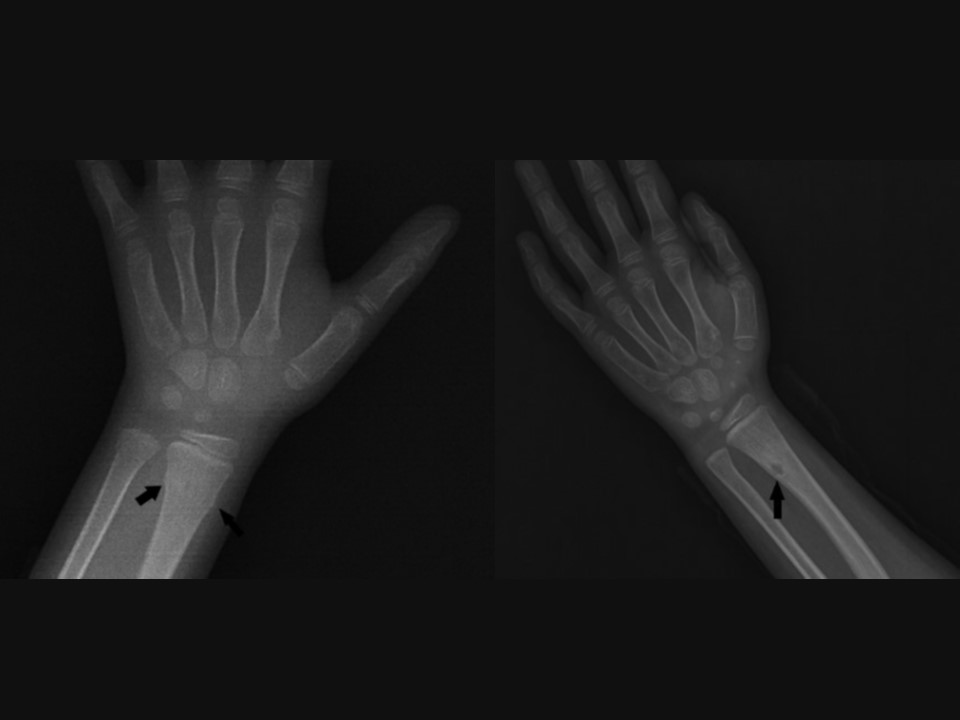
4-year-old girl presented to emergency department with left wrist pain after trauma. Radiographs demonstrated a torus fracture of the distal radius. At the third month following the trauma, a control radiograph is obtained. In the radiograph, a radiolucent lesion close to the former torus fracture site is noticed (Figure 2). Then, CT is performed for further examination (Figure 3). CT demonstrated cortical, well-circumscribed non-expansile subcentimeter lesion.
Downloads
Published
2022-04-26
How to Cite
Karaman, Z. F., & Onem, M. (2022). Pediatric Posttraumatic Cystic Bone Lesion. The Journal of Pediatric Academy, 3(1), 35–36. https://doi.org/10.51271/jpea-2022-171
Issue
Section
Image Corner
License
Copyright (c) 2022 The Journal of Pediatric Academy

This work is licensed under a Creative Commons Attribution-NonCommercial-NoDerivatives 4.0 International License.
The JPA offers users open access to reach all published articles freely within the framework of
NC-ND 4.0)” license.